
Neuromuscular disorders need breakthrough innovation. Now.
Clinical unmet needs are many.
Progressive muscle deterioration remains the hallmark of many neuromuscular disorders.
Myolex continues to advance clinical research.
What if clinicans could…

Peer-reviewed scientific articles showing EIM sensitivity to disease progression and drug effects, reliability, and ease of use
IRB-approved clinical studies by pharmaceutical companies
Issued patents. +2 pending.
Myolex Breakthrough Innovations


Single, quantitative, instant, and more comprehensive measure of muscle composition and structure.

A muscle condition measurement tool that can assist clinical researchers and pharmaceutical scientists in identifying muscle abnormalities significantly early, such as within rapid muscle deterioration conditions, such as with ALS.

The first muscle measurement tool that is accurate, non-invasive, easy-to-use, painless, and portable that enables research subjects to successfully, and more frequently, measure and monitor their muscle condition at home.

Precise, personalized, highly sensitive, and faster muscle condition measurement tool that enables pharmaceutical companies to conduct significantly smaller and faster clinical trials with neuromuscular disorders, such as ALS and DMD.
Capture muscle tissue compositional & micro-structural changes with one comprehensive measure
Including:
- Muscle fiber atrophy and disorganization
- Deposition of fat and connective tissues
- Presence of edema, inflammation
- Other disease pathologies
In ALS, the main feature of muscle diagnostic concern is myofiber atrophy, followed by detecting connective tissue increasing within a muscle. Myolex mScan captures both.
In Duchenne Muscular Dystrophy (DMD), inflammation, fatty deposition, connective tissue, and myofiber hypertrophy and atrophy are all captured within one mScan scanning.

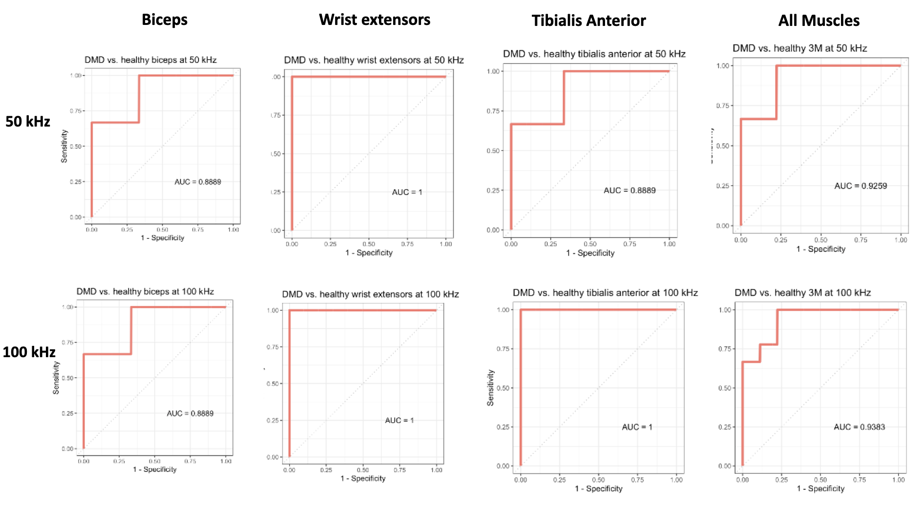
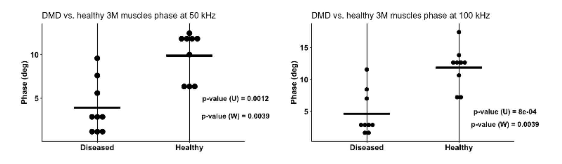
Duchenne muscular dystrophy: Differentiating normal vs. abnormal muscles
Myolex mScan’s EIM-driven precursor (mView) shows a significant difference between healthy and diseased muscles in Duchenne muscular dystrophy (DMD) versus healthy individuals at both 50 and 100 kHz using a 2-group test for matched groups, as shown below (Unpublished data).
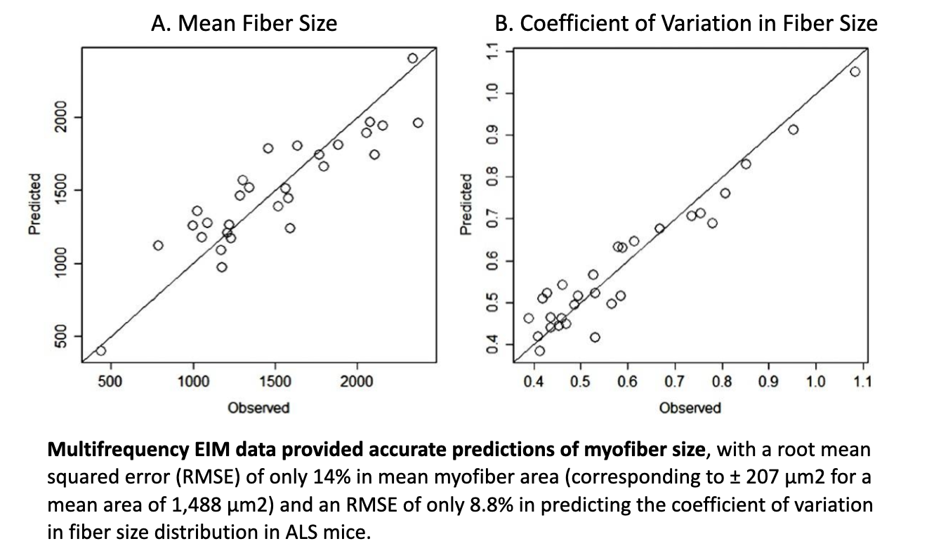
Able to predict myofiber atrophy size, vital to aid diagnosis
EIM has the unique ability to provide virtual biopsy-type data, showing high concordance between observed vs. predicted values.
The Myolex EIM impedance-based approach is very sensitive to deteriorating effects of injury and disease and has demonstrated that it can provide predictive variables to assess myofiber size and distribution with good accuracy, particularly in diseases where myofiber atrophy is the predominant histological feature.
Importantly, this can be done non-invasively, without the requirement for biopsy or burdensome quantification. Because EIM provides spectral data on muscle composition, deep learning techniques, extracting all information buried within the impedance data set, can then be leveraged to optimize EIM’s ability to assess muscle fiber size, contractility, and composition (Ref. 8).
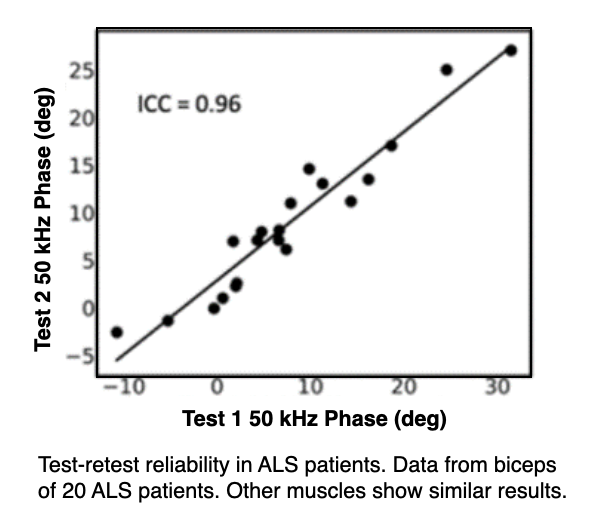
EIM is highly reliable and reproducible
Myolex mScan, produced with support from NIH/NINDS Phase 2B SBIR, also offers excellent reproducibility and reliability across a group of ALS patients. This data was collected by staff at Barrow Neurological Institute with only limited training (Ref. 9).
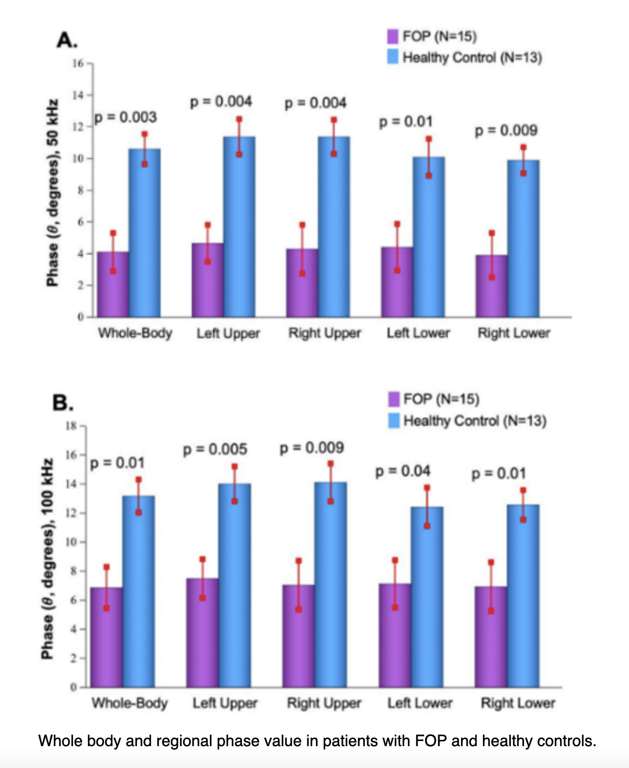
Other rare neuromuscular conditions: muscle integrity measured with EIM at‑home
We aimed to define a novel biomarker in Fibrodysplasia ossificans progressiva (FOP) that enables reliable assessment of musculoskeletal tissue integrity. Considering logistical difficulties that FOP patients often face, our goal was to identify an at-home biomarker technique.
Myolex mScan, along with an accompanying iPad (with EIM data acquisition software), and a BCH-approved Zoom account were shipped to each study participant. Multiple muscle groups were characterized per participant in a 45-min period.
At 50 kHz, EIM was able to differentiate between more severely afflicted muscle regions, as indicated by lower phase value, and regions with healthier unaffected muscle.
This suggests that EIM is capable of detecting regional changes in muscle tissue integrity in FOP (Ref. 10).
For ALS: Opportunity to detect muscle deterioration significantly earlier
Myolex EIM research has shown the ability to identify a decline in the muscle condition, significantly earlier than other measures. Human studies assessing single limb progression of muscle weakness could do the same.
Four technological advances uniquely align that enable Myolex mScan to accurately detect significantly early muscle abnormality (Ref. 11).
What if smaller, faster clinical trials and at-home muscle measurement…
Were a reality?
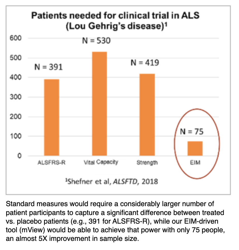
Smaller, faster clinical trials? It's now possible
EIM offers substantial flexibility in terms of its implementation. One can tailor study to a single body region which is most affected by ALS, an approach that cannot be used readily with other disease-tracking tools.
This could translate into tracking a single limb that is most rapidly progressing from the disease during a clinical trial rather than adopting a far less sensitive assessment of whole-body decline.
By performing such directed measurements, it becomes possible to detect a treatment effect over shorter periods of time and with fewer subjects, helping to facilitate and speed the completion of clinical trials.
EIM has the potential to serve as a powerful biomarker in ALS clinical trials that will be able to reduce clinical trial sample size requirements dramatically (Ref. 3).
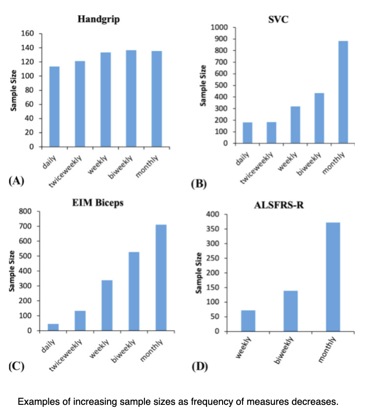
Frequency can reduce clinical sample sizes
The effect of more frequent muscle measurements results in smaller sample size estimates.
For most measures, there is an increase in effect size when more frequent measurements are made. This results in a marked reduction in sample size, in part, because sample size is inversely related to the square of the effect size.
Frequent at-home measurements, using tools such as EIM, holds the prospect of tracking progression more frequently, thereby reducing sample size requirements for clinical trials in ALS while also being acceptable to the patients (Ref. 12).
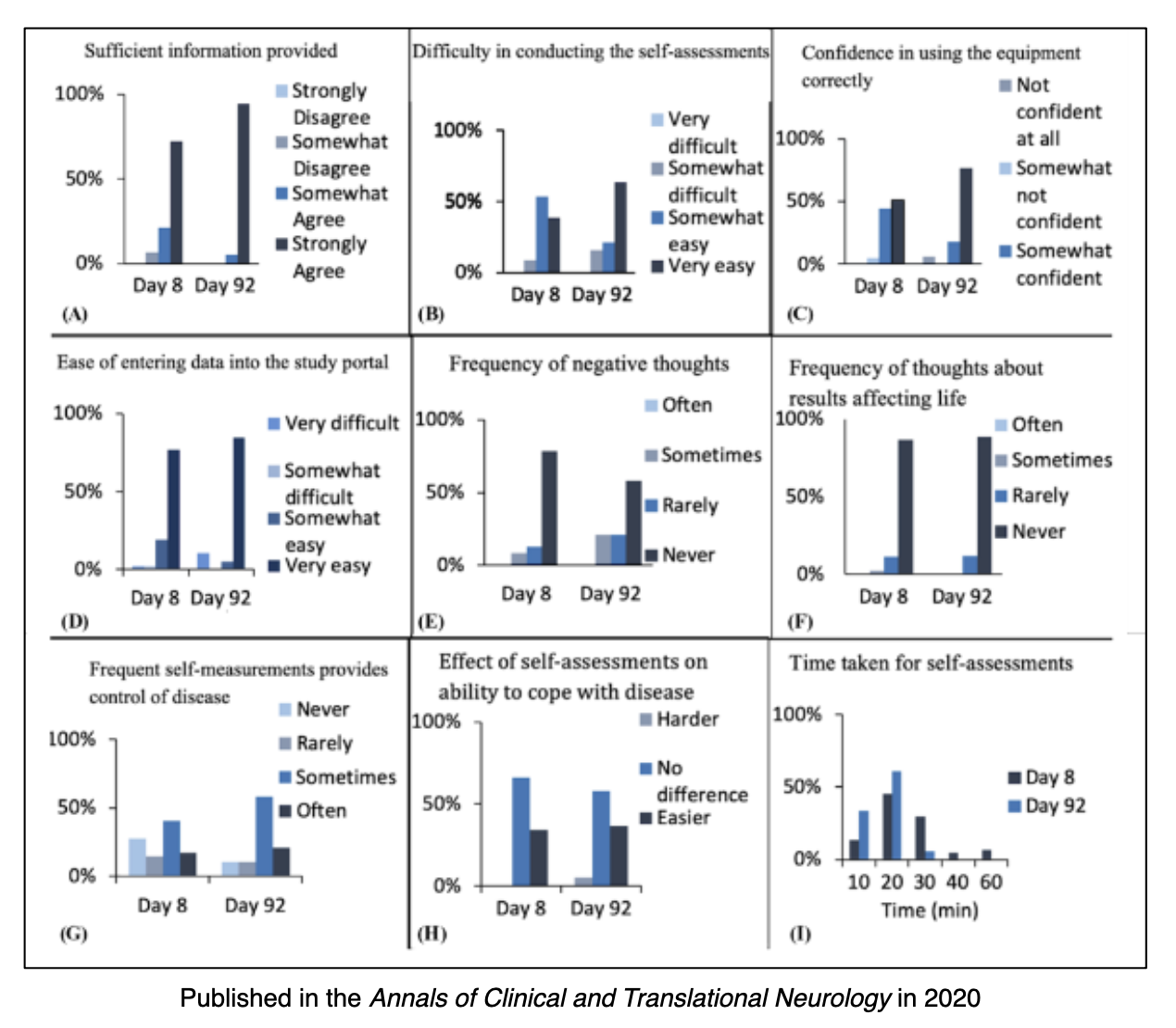
ALS patients can feel more in control at-home
One of the key objectives of this The ALS At Home study was to determine whether participants with ALS or their caregivers could be trained to assess their clinical status by performing a variety of outcome measures at home (Ref. 12).
Conclusions:
- Participants generally felt that they had improved in their ability to take measurements over time, with most completing the entire set of measurements within 20 min by 92 days.
- Of the ALS participants who persisted in measurement, most indicated that the study actually made them feel more in control of their disease.
- The generally positive reactions to home assessments could indicate greater engagement and persistence, as well as a greater sense of personal responsibility.
-
More frequent measurements at-home, greater certainty in ALS progression
By obtaining patient data far more frequently, clinicians will be able to greatly increase their ability to accurately monitor muscle condition change over time (Ref. 13).
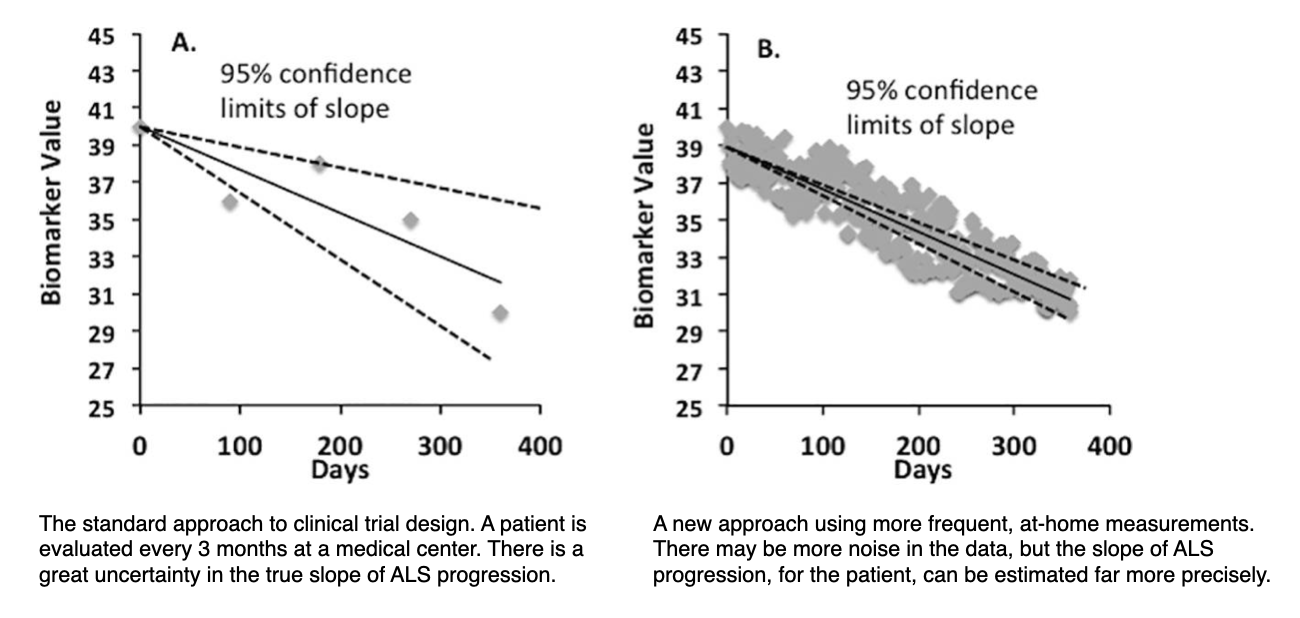
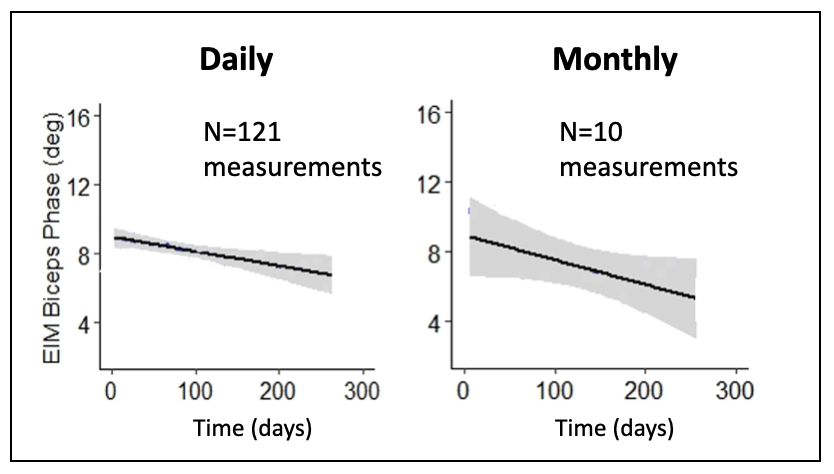
Daily, at-home measurements leads to more accurate tracking
This figure shows data that demonstrates greatly improved accuracy in quantifying the rate of decline in one patient’s biceps. This improved accuracy means better tracking of individual patients and clinical trials with fewer subjects that can be done at home.
Daily, at-home measurements could be performed with a greater 10X improvement over monthly measurements in terms of reductions in sample size (Ref. 5).
Let’s Discover Together.
1309 Beacon Street, Suite 300
Brookline, MA 02446
Phone: 888.382.8824
info@myolex.com
